Diagram Of Bones In Neck And Shoulder - Human Appendicular Skeleton Biology For Majors Ii. Shoulder girdle , radiographs : Related posts of bones of the head neck and shoulder. 7 draw labelled diagram showing the relations of shoulder joint. These consist of the arm, located between the shoulder and elbow joints; Joints hold your bones together and allow your rigid ball and socket joints, like your hip and shoulder joints, are the most mobile type of joint in the human body.
The shoulder joint is one of the most mobile in the body, at the expense of stability. This bone is normally cylindrical but flares when it comes near the elbow and becomes flat. A diagram of the human skeleton showing bone and cartilage. The shoulder bones can easily be affected by falls or accidents, in addition to arthritis. It is a long bone that acts as a rod between the shoulder blade (scapula) and the sternum (breastbone) to link them.
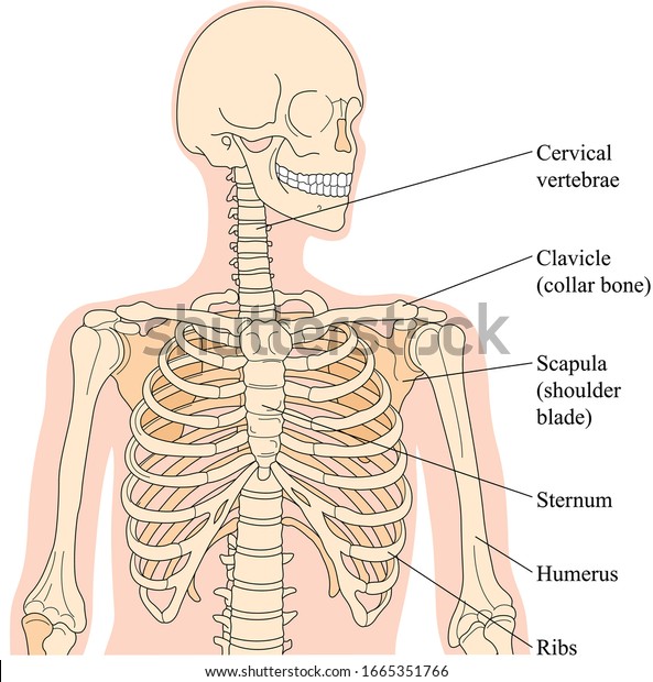
Bones Neck Chest Shoulder Girdle Stock Vector Royalty Free 1665351766 from image.shutterstock.com Large,flat, triangular bone of the shoulder. The compact bone is the smooth and very hard part of the bone. In adults the long bones of the legs and arms are filled with yellow marrow. The shoulder joint is one of the most mobile in the body, at the expense of stability. Muscles and bones illustration for kids. Here, we shall consider the factors the permit movement, and those that contribute towards joint structure. These consist of the arm, located between the shoulder and elbow joints; It's a long, thin bone that curves outward at the middle of your body and curves inward on the end where it goes to the shoulder.
Collarbone.bone that joins the sternum and scapula.
Muscles and bones illustration for kids. Bones in the hand and wrist right hand. Femoral neck fractures or fractures of the neck of the femur are fractures of the proximal femur in the region between head of femur and intertrochanteric line. The femur is the largest bone in the body and the only bone of the thigh (femoral) region. The shoulder joint is one of the most movable joints in the human body. In adults the long bones of the legs and arms are filled with yellow marrow. What are the bones called in your neck shoulder area and upper back socratic. The shoulder joint is one of the most mobile in the body, at the expense of stability. Here, we shall consider the factors the permit movement, and those that contribute towards joint structure. The upper limb is divided into three regions. Located on the lateral side of the proximal humerus is an expanded. Bones of the face hyoid in the neck, below the tongue (held in place by ligaments and muscles between it and the styloid process of the temporal bone). The bony structures of the shoulder include the pectoral girdle and one arm bone.
It is a long bone that acts as a rod between the shoulder blade (scapula) and the sternum (breastbone) to link them. The shoulder bones consists of two bones : In adults the long bones of the legs and arms are filled with yellow marrow. Webmd's shoulder anatomy page provides an image of the parts of the shoulder and describes its function, shoulder problems, and more. A diagram of the human skeleton showing bone and cartilage.
Human Shoulder Anatomy Koibana Info Neck And Shoulder Muscles Shoulder Muscle Anatomy Muscle Anatomy from i.pinimg.com It allows the upper limb to have a wide array of movements. All of your bones, except for one (the hyoid bone in your neck), form a joint with another bone. Femoral neck fractures or fractures of the neck of the femur are fractures of the proximal femur in the region between head of femur and intertrochanteric line. The upper limb is divided into three regions. These consist of the arm, located between the shoulder and elbow joints; The seven bones of the top part of the vertebral column, located in the neck region. Boneka stitch jumbo warna biru. There are five major shoulder bones.
The axial skeleton and the appendicular it forms the ball and socket joint of the shoulder with the scapula and forms the elbow joint with the lower arm bones.
Each arm is attached to a shoulder blade or scapula (say: It is made up of bones. Each arm is attached to a shoulder blade. Muscles and bones illustration for kids. Collarbone is the only long bone in our body that lies horizontally. There are 33 bones in the spine. The clavicle, or collarbone, lies horizontally at the root of the neck. The axial skeleton and the appendicular it forms the ball and socket joint of the shoulder with the scapula and forms the elbow joint with the lower arm bones. (4) just below the neck, there are shoulder bones on both sides of the skeleton. The bony structures of the shoulder include the pectoral girdle and one arm bone. Related posts of bones of the head neck and shoulder. Very soon we'll move on to muscles! Collar bones and shoulder there is also a small bones called breast bone in the chest region, in front of our body.
The compact bone is the smooth and very hard part of the bone. (4) just below the neck, there are shoulder bones on both sides of the skeleton. Shoulder joint of human body anatomy infographic diagram with all parts including bones ligaments muscles bursa cavity capsule cartilage membrane for medical science education and health care. The seven bones of the top part of the vertebral column, located in the neck region. 2.1 bones of the shoulder girdle 2.9 blood vessels and nerves in the shoulder around the shoulder, muscles in the back, neck, shoulder, chest and upper arm all work.
Human Upper Limb Shoulder Girdle Arm And Hand Showing Bones And Download Scientific Diagram from www.researchgate.net The shoulder joint is one of the most movable joints in the human body. The seven bones of the top part of the vertebral column, located in the neck region. Each arm is attached to a shoulder blade. Examples include cranial bones (protecting the brain), the sternum and ribs (protecting the organs in the thorax), and the scapulae (shoulder blades). Large,flat, triangular bone of the shoulder. These consist of the arm, located between the shoulder and elbow joints; On series you can directly access the radiological images of the. All of your bones, except for one (the hyoid bone in your neck), form a joint with another bone.
The bones of the shoulder consist of the humerus (the upper arm bone), the scapula (the shoulder it forms the front portion of the shoulder girdle and is palpable along its entire length with a gentle the top end of the humerus consists of the head, the neck, the greater and lesser tubercles, and the.
The compact bone is the smooth and very hard part of the bone. Collarbone.bone that joins the sternum and scapula. All of your bones, except for one (the hyoid bone in your neck), form a joint with another bone. The structure of bone with diagram and definitions. Types of bones with examples. Each arm is attached to a shoulder blade or scapula (say: Examples include cranial bones (protecting the brain), the sternum and ribs (protecting the organs in the thorax), and the scapulae (shoulder blades). Collar bones and shoulder there is also a small bones called breast bone in the chest region, in front of our body. Other important bones in the shoulder include On series you can directly access the radiological images of the. The shoulder and arm bones can be broken or dislocated by traumatic injuries. Webmd's shoulder anatomy page provides an image of the parts of the shoulder and describes its function, shoulder problems, and more. The bony structures of the shoulder include the pectoral girdle and one arm bone.
Share :
Post a Comment
for "Diagram Of Bones In Neck And Shoulder - Human Appendicular Skeleton Biology For Majors Ii"


Post a Comment for "Diagram Of Bones In Neck And Shoulder - Human Appendicular Skeleton Biology For Majors Ii"